
Images from the National Museum of Health and Medicine

These photographs were taken at The National Museum of Health and Medicine on the grounds of Walter Reed Army Medical Center in Washington DC. Since that time, Walter Reed has closed and the museum has moved to a new, but smaller, site in Silver Springs MD.

Hanaoka Seishu's Surgical Casebook
“A Surgical Casebook” is a manuscript of hand-painted pictures commissioned by Hanaoka Seishu, a pioneering Japanese surgeon who was the first to use general anesthesia to remove tumors from cancer patients. The colorful, often charming, pictures in this casebook capture the likenesses of the men and women who came to Hanaoka for treatment; and, importantly, they depict, quite graphically, the medical and surgical problem to be treated. Clearly, Hanaoka engaged an artist of considerable talent to make a visual document of the patient’s condition before surgery. This suggests a local artist or an artistically-talented medical associate, because Hanaoka would have encountered the patients depicted in this book over a period of many years. The labels attached to the pictures provide information about a patient’s place of residence, occasionally his or her name and livelihood, and sometimes a diagnosis.

Hanaoka Seishu’s fame is based on his invention of an oral anesthesia that could render a patient unconscious for long enough to allow him to remove deep tumors. Hanaoka was born to a physician’s family in Kii Province (today’s Wakayama Prefecture), a remote, mountainous region of south central Japan, in 1760. At age twenty-two he went to Kyoto, where he studied both traditional Chinese-style medicine and Western-style surgical techniques; at age twenty-five he took over the family business and began to practice an eclectic style of medicine that combined these two traditions. He was greatly concerned with his inability to treat cancer patients, and over a period of twenty years he developed an herbal concoction he called ‘mafutsusan.’ It was made up of several highly toxic plants, including Korean asagao, Japanese aconite, Chinese angelica, and Arisaema japonicum, among others. It did not include opium derivatives which were only beginning to be identified by European doctors. The herbs were ground into a paste, boiled with water, and administered to the patient by mouth well before the surgery. The narcotic effects of this anesthetic could last as long as 24 hours, allowing him to surgically remove many different kinds of tumors which previously had been inoperable (Access the book).
Epilepticus sic curabitur
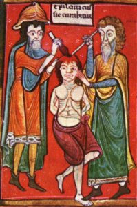 The surgical treatment of epilepsy is not a recent innovation. As early as ancient Greek and Roman times, and in the Middle Ages, trepanation (the opening of the skull) was occasionally carried out on people with epilepsy. There were many different reasons for doing this, however. There was seldom a rational, medical reason for the operation, for instance to raise a piece of the skull cap which had entered the inner part of the skull as a result of an injury, perhaps in war, and which was thought to cause epileptic seizures in the patient. In such a case, the surgeon tried to raise the piece of bone and thus remove the cause of the epilepsy. Mostly, however, there were mystical, superstitious reasons for the trepanation operations which were performed. It was thought that opening the skull cap would cause the demons of the sickness, poisonous gases or disease-causing juices to escape. The practice of cauterisation was based on the same beliefs. In this picture, the person with epilepsy ('epilepticus') is undergoing both procedures - trepanation and cauterisation - at the same time.
 Epilepticus sic curabitur ('The way to cure an epileptic') Sloane Manuscript, collection of medical manuscripts, end of the 12th century - British Museum, London
Phrenology
 Phrenology (from Greek: φρήν, phrēn, "mind"; and λόγος, logos, "knowledge") is a defunct field of study, once considered a science, by which the personality traits of a person were determined by "reading" bumps and fissures in the skull. Developed by German physician Franz Joseph Gall around 1800, the discipline was very popular in the 19th century. In 1843, François Magendie referred to phrenology as "a pseudo-science of the present day."[1] Phrenological thinking was, however, influential in 19th-century psychiatry and modern neuroscience.[2]
Eponims in Neuroanatomy
Herein it is reviewed several contributions to neuroanatomy in the period extending from the renaissance, when interest in the old Greek medicine revived, to the end of the 18 th century. I will concentrate on three eponyms: 1) the circle of Willis; 2) the Sylvian fissure; 3) the foramen of Monro. The work of these three eponymists will be put into the context of the evolution in neuroanatomy of the period in which they worked. Contributions to neuroanatomy have been added over the centuries, although to a lesser degree in the period between Galen (130-200) and the renaissance. Galen’s work had been authoritative for over twelve centuries. Medieval physicians kept to his anatomy unquestioning. It had about the same status in medicine as the bible in religion. Important renovations started in the renaissance period.
Subscribe to:
Posts (Atom)